
Figure 1.
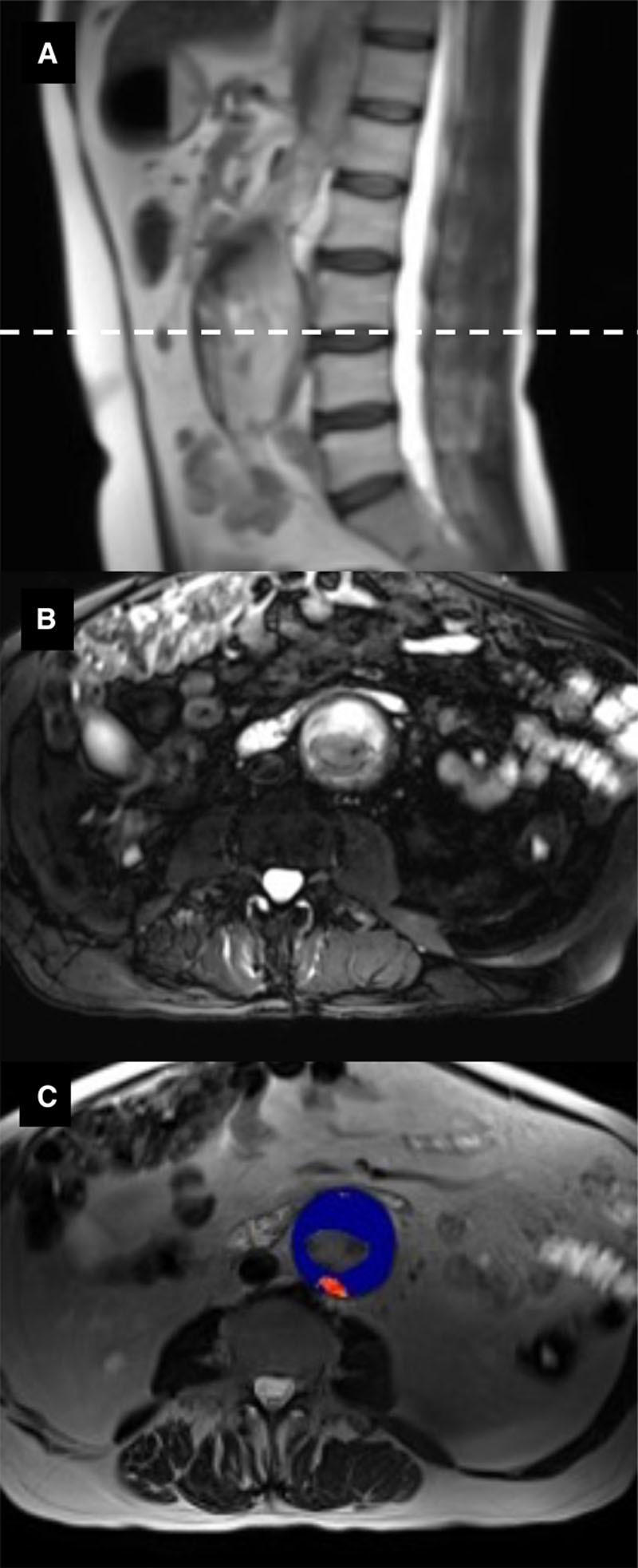
MRI of abdominal aortic aneurysm. A, T2-weighted HASTE (Half Fourier Acquisition Single Shot Turbo Spin Echo) sequence in the sagittal plane. B, Cross-sectional image (dashed line in A) using a T2-weighted fat-saturated sequence to highlight intraluminal thrombus (white) within the aneurysm. C, T2* map (blue) overlying the T2-weighted HASTE sequence (B), demonstrating enhancement of the posterior aneurysm wall with ultrasmall superparamagnetic particles of iron oxide (USPIO) (red).