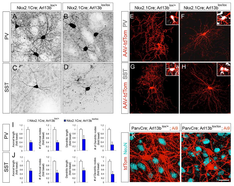
Figure 1. Deletion of Arl13b in interneurons results in morphological defects.
(A–B) Striatal PV+ interneurons were labeled with anti-PV antibodies in Nkx2.1Cre; Arl13blox/+ (A) and Nkx2.1Cre; Arl13blox/lox (B) brains. (C, D) Striatal SST+ INs were labeled with anti-SST antibody in Nkx2.1Cre; Arl13blox/+ (C) and Nkx2.1 Cre; Arl13blox/lox (D) brains. (E–H) Representative images of PV+ (E, F) or SST+ INs (G, H) interneurons from AAV2-FLEX-tdTomato injected Nkx2.1Cre; Arl13blox/+ (E, G) and Nkx2.1Cre; Arl13blox/lox (F, H) brains. Insets (E–H) show co-labeling of tdTom+ neurons with PV (E, F) and SST (G, H) antibodies. (I–J) Quantification of morphological defects of PV+ (I) and SST+ (J) INs in Nkx2.1Cre; Arl13blox/lox brains [P30]. Data shown are mean ± SEM. *P<0.05 (Student’s t-test;). (K–L) Representative images of tdTom+ INs from ParvCre; Arl13blox/+; Ai9 (K) and ParvCre; Arl13blox/lox; Ai9 (L) brains [P60]. Neurons were co-labeled with anti-NeuN antibodies. Data shown are mean ± SEM. *P<0.05 (Student’s t-test, p[PV+ axonal length] = 5.32281E-06, p[PV+ axonal node] = 0.0005, p[PV+ dendritic length] = 0.0001, p[PV+ dendritic node] = 0.0003, p[SST+ axonal length] = 7.92531E-06, p[SST+ axonal node] = 0.0002, p[SST+ dendritic length] = 0.002, p[SST+ dendritic node] = 0.0003). 42 cells from 4 different brains were analyzed per group. Scale bar, 25μm (A–D); 50μm (E–H); 20μm (K, L). See also Figure S1, Figure S2, and Figure S3.

