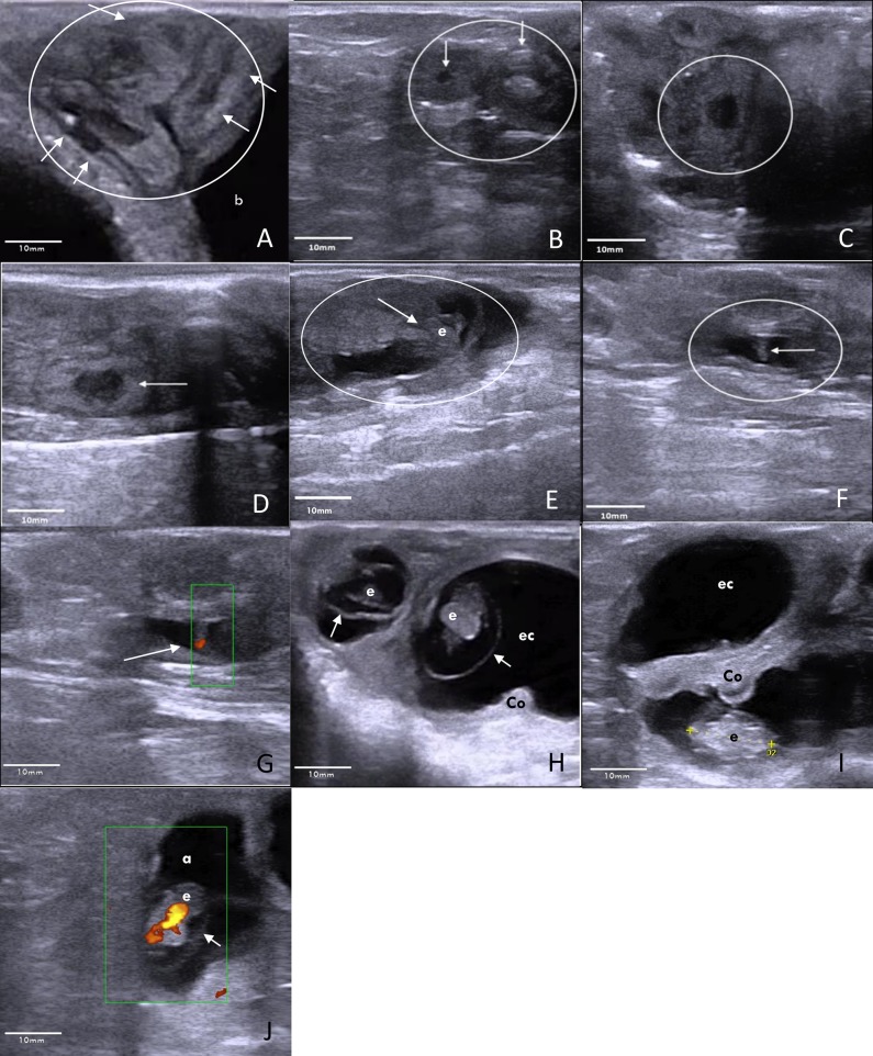
Fig 1. Transrectal ultrasound (Esaote Germany, MyLab™One VET, equipped with electronic linear array 10–5 MHz transducer (SV3513 Vet)) images of the ovary, uterus and developing conceptus during early pregnancy in sheep (oestrus = day 0).
A Day 10: The coiled uterus (circle) is located cranial to the bladder (b). Within the coiled uterine horns, the lower echogenic endometrium (arrows) is well distinguished. B Day 10: Two corpora lutea (arrows) are present on the ovary (circle), documenting recent ovulation. C Day 12: Trophoblastic expansion (within the circle): At the site of trophoblastic expansion the endometrium appeared enlarged. A dark oval shaped structure expanded within the uterine lumen. Boundaries between endometrium and trophoblast could not be distinguished at this stage. Yet the trophoblast expanded the uterine lumen exceeding the regular endometrial height by 2-fold. D Day 14: Trophoblastic expansion (arrow): Low echogenic oval-shaped trophoblast within the uterine lumen, further expands in length but not in height. E Day 17: Embryonic vesicle (circle) with embryo (arrow) within. F Day 19: Embryo (arrow), distinctly present inside the embryonic vesicle (circle). G Day 19: Power Doppler of the embryo (arrow) heartbeat. H Day 29: Two embryo’s (e) surrounded by the amniotic membranes (arrows). A cotyledon is in close proximity of the embryo (Co). Crown rump length of the embryo measures 14 mm. Fluids of the embryonic cavity (ec) expand further into the adjacent uterine horn. I Day 29: Embryo (e), situated below a cotyledon (Co). Above this the fluid filled embryonic cavity (ec), which expands into the uterine horn. J Day 29: Power Doppler of the embryo heart. Embryo (e) is surrounded by the amnion (a) and the fluid filled embryonic cavity (arrow).

