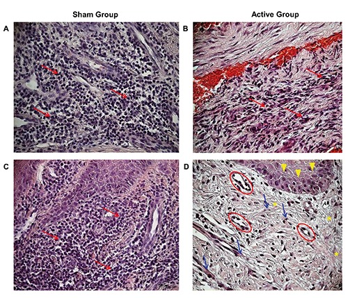
Figure 3.

Hematoxylin and Eosin staining of DFUs at 20X magnification. Sham Group at baseline (A) and after the treatment with non-functioning TMR® device (C): Red arrows indicate granulocytes and macrophages. Active Group at baseline (B) and after the treatment with functioning TMR® device (D): yellow head arrows indicate a layer of cuboidal epithelial cells, red circles point to endothelial cells, blue arrows indicate fibroblasts, and yellow asterisks point to collagen fibers.
