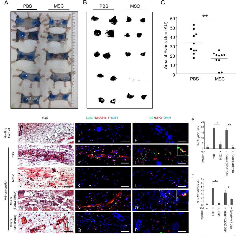
Figure 4.
MSCs reduce vessel destruction and hemorrhage in immune complex-mediated vasculitis. (A): 100 µl PBS or 2.5 × 10⁵ AT-MSCs were intradermally injected to both sides of shaved dorsal skin of mice. The Arthus reaction was elicited by i.v. injection of 100 µl PBS solution containing 2% BSA and 1% Evans blue. Afterward anti-BSA antibody was intradermally injected to the area that had been injected with AT-MSCs or PBS. Four hours later, mice were sacrificed and skin specimen were harvested and digitally photographed. (B– C): The areas of Evans blue spots on skin were analyzed by Image J. n = 10; **, p < .01 by Mann-Whitney tests. (D–T): Mice were intra-dermally injected with PBS or nontransfected MSCs or SOD3-siRNA transfected MSCs or control-siRNA transfected MSCs and subjected to Arthus reaction. Mice received rabbit IgG served as healthy controls. Representative pictures of paraffin-embedded skin sections from 4 mice of each group with H&E staining (D, G, J, M, P), immunostaining of murine neutrophil marker Ly6G (green) and NETs marker DNA/Histone-1 (red) (E, H, K, N, Q), and immunostaining of NE (green) and MPO (red) (F, I, L, O, R) are shown. Nuclei were counterstained with DAPI (blue) in immunostainings. White arrows in H&E staining indicate blood vessels. Scale bar: 50 µm. (S–T) The depicted are mean ± SEM of Ly6G+ cells (S) and NE+ MPO+ cells (T) counted from each section of 4 mice. n = 4; *, p < .05; **, p < .01 by Mann-Whitney tests. Abbreviations: AU, arbitrary unit; MSCs, mesenchymal stem cells; MPO, myeloperoxidase; NE, neutrophil elastase.

