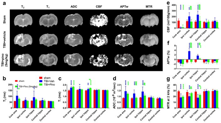
Fig. 3.
Quantitative multiparametric MRI assessment of neuroprotection by pinocembrin in rats with traumatic brain injury (TBI). (a) Representative images of multiparametric MRI signals at 3 days post-TBI in sham, TBI + vehicle, and TBI + pinocembrin (pino; 5 mg/kg) groups (n = 5 per group). Notably, the TBI lesion became heterogeneous with areas of high and low APTw signal intensities. The APTw signal intensity of the perilesional region increased dramatically in the vehicle-treated TBI rats, but less so in the pinocembrin-treated TBI rats. The ADC, which probes the diffusion rate of water molecules, had a significantly lower signal in the pinocembrin-treated TBI rats than in the vehicle-treated TBI rats. The CBF signal showed a similar trend with pinocembrin treatment. (b–g) Quantitative analysis of multiparametric MRI signal intensities in five regions of interest (ROIs). In general, the ADC, CBF, and APTw MRI signals in surrounding areas (ipsilateral cortex and hippocampus) showed significant increases in the vehicle-treated TBI group that were ameliorated in the pinocembrin-treated TBI group. Conversely, the MTR showed a significant decrease in the vehicle-treated TBI group that was reversed by pinocembrin treatment. *P < 0.05, **P < 0.01, ***P < 0.001, ****P < 0.0001 compared with the sham group; #P < 0.05, ##P < 0.01, ###P < 0.001, ####P < 0.0001 compared with the vehicle-treated TBI group.

