Fig. 4.
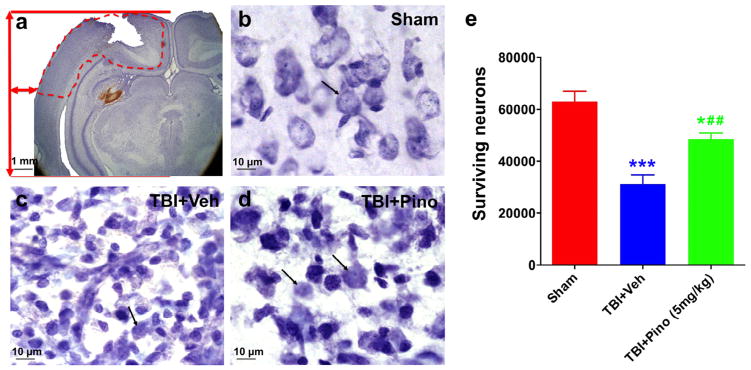
Cresyl violet staining and stereological quantification of surviving neurons at 3 days after traumatic brain injury (TBI) in rats. (a) Representative Cresyl violet-stained brain sections from rats after TBI. The location in the ipsilateral cortex used to quantify the number of surviving neurons by stereology is delineated by a dashed red line. (b–d) Morphologic changes in cortical neurons after TBI. Sham brain showed healthy neurons, with large nuclei and distinct nucleoli, indicated by black arrows (b). TBI brain showed profound neuronal degeneration as indicated by the neuronal shrinkage and pyknotic nuclei (c). With pinocembrin treatment, many neurons exhibited normal appearance, but shrunken cells with pyknotic nuclei were still evident (d). (e) Volumetric density of viable neurons assessed in ipsilateral cortex by unbiased stereology (n = 5 per group). The TBI + vehicle group showed a significant decrease in viable neurons, compared with the sham group (***P < 0.001). Importantly, rats treated with pinocembrin had significantly more viable neurons than did those treated with vehicle (##P < 0.01), although the number was still less than that of the sham group (*P < 0.05). Scale bar in (a), 1 mm; scale bar in (b–d): 10 μm.

