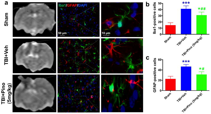
Fig. 5.
Glial response in the perilesional hyperintense brain areas at 3 days after traumatic brain injury (TBI) in rats. (a) APTw images and immunofluorescence labeling of microglia (Iba1, green) and astrocytes (GFAP, red) in rat brains from the sham, TBI + vehicle, and TBI + pinocembrin groups at 3 days post-TBI. Consistent with the changes to APTw signal in the perilesional region, at 3 days after TBI, fluorescence intensities of Iba1 and GFAP were significantly higher in the vehicle-treated TBI group than in the sham group. However, pinocembrin moderated the level of increase. (b, c) Quantitative analysis of Iba1-positive reactive microglia and GFAP-positive reactive astrocytes in the hyperintense surrounding areas (n = 5 per group). The TBI + vehicle group had greater numbers of Iba1- and GFAP-positive reactive cells than did the sham group (***P < 0.001). The TBI + pinocembrin group had significantly fewer Iba1- and GFAP-positive reactive cells than did the TBI + vehicle group (#P < 0.05, ##P < 0.01), but still had more than the sham group (*P < 0.05). The data suggest that changes in the APTw signals may be associated with microglial and astrocyte activation. Scale bars in (a) middle column, 50 μm; right column, 10 μm.

