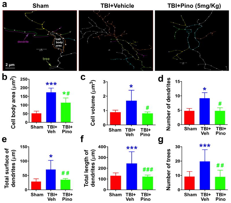
Fig. 6.
Morphometric assessment of microglia in the APTw-hyperintense areas of sham, TBI + vehicle, and TBI + pinocembrin groups (n = 5 per group). We used Neurolucida software, a computer-based tracing system, to trace and analyze the structure (dendrites and cell bodies) of microglia under a 40 × objective. (a) The traced images. (b–g) Changes in cell body area, cell volume, number of dendrites and trees, and total surface and length of dendrites. Cell body area of the microglia was greater in the TBI + vehicle group than in the sham group. Although, microglia in the TBI + pinocembrin group had significantly smaller cell bodies than microglia in the TBI + vehicle group, they remained larger than those in the sham group. Similarly, microglia in the TBI + vehicle group exhibited greater cell volume and total surface of dendrites, greater numbers of dendrites and trees, and longer dendrites than those of the sham groups, and pinocembrin treatment reversed these changes. Scale bars in (a): 2 μm. *P < 0.05, ***P < 0.001 vs. sham; #P < 0.05 vs. TBI + vehicle.

