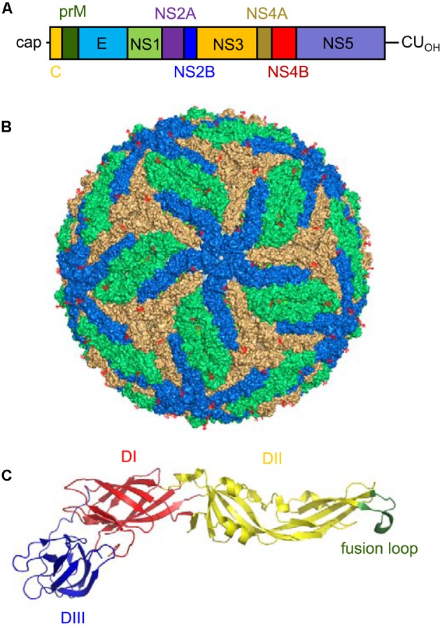
FIGURE 3.

Genomic organization, virion structure, and organization of the E glycoprotein of Zika virus (ZIKV). (A) Schematic view of the genomic organization of ZIKV. The single open reading frame (ORF; boxes) that encodes both structural (C-prM/M and E) and non-structural (NS) proteins (NS1, NS2A, NS2B, NS3, NS4A, NS4B, and NS5) is flanked by two untranslated regions (UTRs). Notice the presence of a 5′ cap and the lack of a poly(A) tail at the 3′ end of the genome. (B) Surface representation of a ZIKV mature particle. The E monomers are colored in blue, orange and green to facilitate the interpretation of their distribution. Image was produced using the cryo-electron microscopy data available (Protein Data Bank entry 5IRE). (C) Structure of a monomer of the soluble ectodomain of E glycoprotein of ZIKV. The ribbon diagram was based on the atomic coordinates solved by X-ray crystallography (Protein Data Bank entry 5JHM). DI in red, DII in yellow, and DIII in blue. Fusion loop is highlighted in green.
