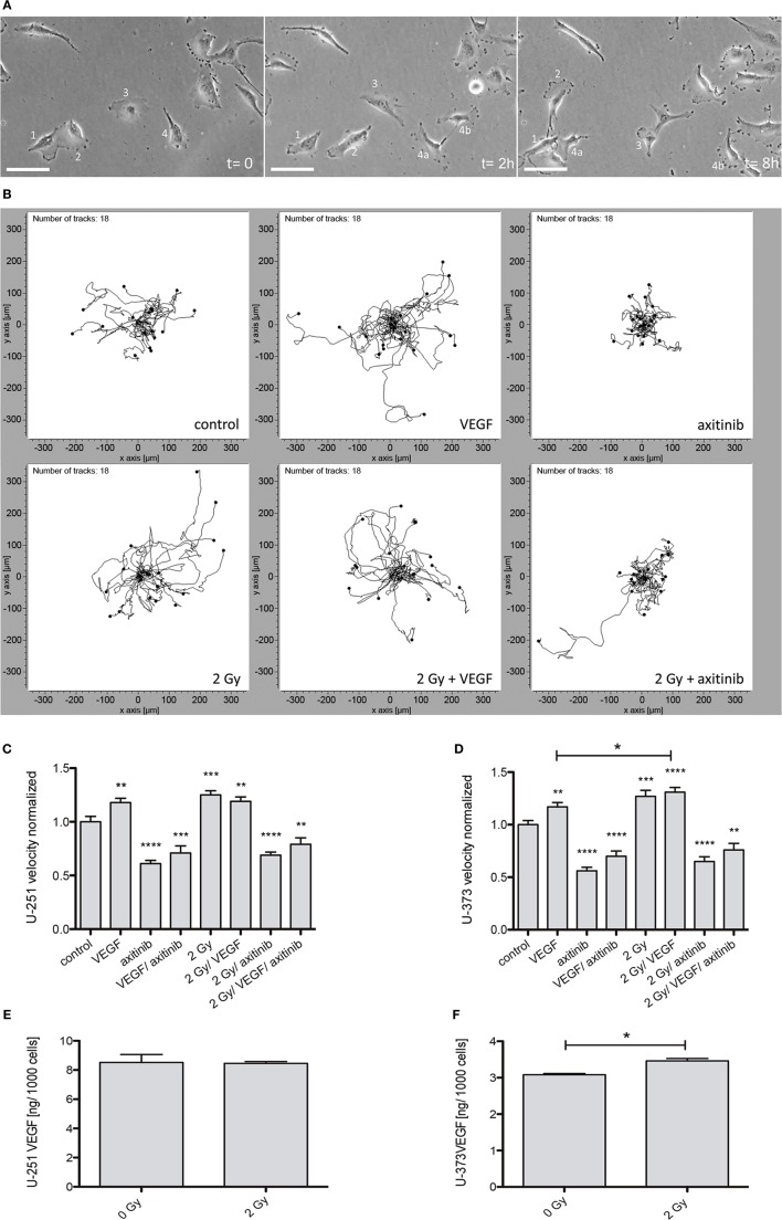
Figure 2.
Influence of vascular endothelial growth factor (VEGF), axitinib, or irradiation with 2 Gy photons on the motility of glioblastoma multiforme cell lines U-251 and U-373. The motility of the cells was analyzed by time-laps videography. Cells were tracked and analyzed with the ibidi chemotaxis and migration tool. (A) Examples from an image stack of Videography. Some cells migrate fast (2), some slowly (3), some even do not migrate (1). Dividing cells (4) often keep contact over longer periods of time (4a, 4b). Scale bars: 50 µm. (B) Migration of U-373 glioblastoma cells under varied conditions. Depicted are the tracked cells in representative fields of view. The software merges all starting points in the origin to get an explicit view of the paths migrated by the cells. It is clearly visible that the migration is undirected (an advantage of videography over other methods to analyze migration). It is notable that some irradiated cells are able to escape the inhibition by axitinib. (C,D) The motility of U-251 and U-373 cells is increased by VEGF as well as by irradiation. In U-373, a combination of both leads to a significant increase in velocity compared to VEGF alone (D), in U-251 no additive effects could be observed. In contrast, axitinib diminishes the motility of untreated cells and the elevated motility after irradiation as well. VEGF and axitinib were added in concentrations of 0.1 and 10 µg/ml, respectively. (E,F) 24 h after 2 Gy irradiation the amount of VEGF in the supernatant of U-251 and U-373 was analyzed. In U-251 there were no significant changes detectable, whereas in U-373 cells VEGF was significantly increased (F). Data are shown as mean ± SEM. Data were tested for significance using one-way ANOVA with Bonferroni multiple comparison post-test. Significant differences are indicated by *p < 0.05; **p < 0.01; ***p < 0.001; ****p < 0.0001.

