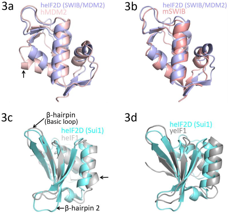
Figure 3.
Superposition of the SWIB/MDM2 domain of heIF2D with (a) N-terminal domain (E25-V109) of human MDM2 (PDB: 1YCR) and (b) SWIB domain of mouse BAF60a (PDB: 1UHR). The arrow in 3(a) points toward helix α4 that shifts by 6.7 Å. Superposition of the SUI1 domain of heIF2D with (c) human eIF1 (heIF1) (PDB: 2IF1) and (d) yeast eIF1 (yeIF1) (PDB: 2OGH). The arrow in 3(c) points toward helix α2 that shifts by 3.0 Å. The structures were superposed using COOT and the figure was made in Pymol.

