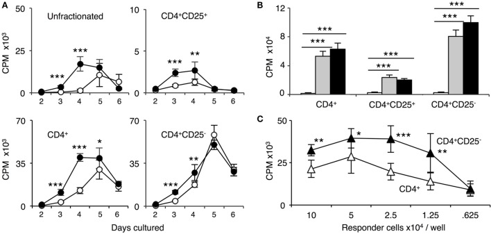
Figure 3.
Comparison of proliferation of naïve unfractionated, CD4+ T cells and CD4+ T cell subsets in mixed lymphocyte culture (MLC) to self-DA, and allogeneic stimulators Piebald Virol Glaxo rat strain (PVG) and Lewis. Proliferation of unfractionated and CD4+, CD4+CD25−, and CD4+CD25+ T cells from naïve DA rats in MLC was assessed. The response to fully allogeneic PVG stimulator cells was compared to self-DA (A,B) and other fully allogeneic Lewis stimulator cells (B). Significant differences are shown as *p < 0.05, **p < 0.01, and ***p < 0.001. (A) Time course with unfractionated cells and CD4+ T cells showed peak proliferation to PVG (●) at day 4 and maintained at day 5, after which it waned. There was significant proliferation to DA (○), peaking at day 5. With CD4+CD25− T cells, proliferation was greatest at day 4 and day 5; however, there was little difference in response to PVG and self-DA. Proliferation of CD4+CD25− T cells was greater than unfractionated and enriched CD4+ T cells. CD4+CD25+ T cells proliferation was much less than that of CD4+ or CD4+CD25− T cells. There was a detectable response to PVG at days 3 and 4 that waned by day 5 and 6. In subsequent experiments, proliferation of CD4+CD25+ T cells was assayed at day 4, and of CD4+ and CD4+CD25− at day 4 or 5. (B) Comparison of proliferation in MLC of CD4+ T cells and subsets to self-DA, PVG, and Lewis stimulator cells. All populations had similar responses to PVG ( ) and Lewis (◼) that were greater than proliferation to self-DA (◻) (***p < 0.001). (C) Comparison of responses of serially diluted naïve DA CD4+ T cells (△) and CD4+CD25− T cells (▲) in MLC stimulated by a constant number of PVG cells. CD4+CD25− T cells had significantly higher proliferation to PVG compared to CD4+ T cells indicating that removal of naïve CD4+CD25+ T cells significantly enhanced proliferation.

