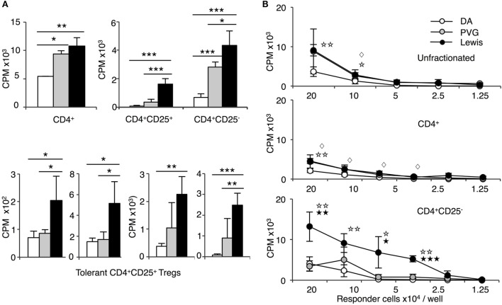
Figure 4.
Comparison of proliferation of CD4+, CD4+CD25−, and CD4+CD25+ T cells from tolerant hosts in mixed lymphocyte culture (MLC) to self-DA, specific-donor Piebald Virol Glaxo rat strain (PVG), and to third-party Lewis. Proliferation of CD4+, CD4+CD25−, and CD4+CD25+ T cells subjected to MLC with either self (DA), specific-donor (PVG), or third party (Lewis) at day 4, assessed by 3H thymidine incorporation and expressed as cpm. (A) Proliferation of cell subsets to donor antigens in MLC. CD4+ T cells from tolerant hosts (top left panel) had a similar response to specific-donor PVG ( ) and third-party Lewis (◼) that was much greater than to self-DA (◻). Two other experiments had similar results. For CD4+CD25− T cells from tolerant hosts (top right panel), the response to specific-donor PVG was less than to third-party Lewis. This response of CD4+CD25− T cells was not greater than that of CD4+ T cells. CD4+CD25+ T cells from DA rats tolerant to PVG graft did not respond to specific-donor PVG, and this response was not greater than to self-DA (top right panel). CD4+CD25+ T cells from tolerant hosts had significantly higher proliferation to third-party Lewis compared to specific-donor PVG or self-DA. Bottom row panel shows 4 of the 8 replicate experiments that confirmed CD4+CD25+ T cells from tolerant hosts did not respond or had low response to specific-donor strain PVG compared to their response to third-party Lewis. Significant differences in responses are marked as * if p < 0.05; ** if p < 0.01, and *** if p < 0.001. (B) Limiting dilution assay of cells from tolerant hosts in MLC. The response of unfractionated cells (top panel) and CD4+ T cells (middle panel) was similar to specific-donor PVG (
 ) and third party Lewis (●) at all points, and was greater than that to self-DA (○). CD4+CD25− T cells from tolerant hosts (bottom panel), in contrast to CD4+ T cells (middle panel), had a marked increased response to third-party Lewis but not to specific-donor PVG. Significant at three dilutions. Collectively, these findings suggest that CD4+CD25+ T cells from tolerant animals paradoxically did not suppress the response of CD4+CD25− T cells from tolerant hosts to specific-donor alloantigen, but retain the capacity to suppress responses to self-DA cells and third-party Lewis alloantigen. Symbols for significant differences are: ⋄ PVG vs DA; ☆ Lewis vs DA; ★ Lew vs PVG; 1 symbol if p < 0.05; 2 symbols if p < 0.01, and 3 symbols if p < 0.001.

