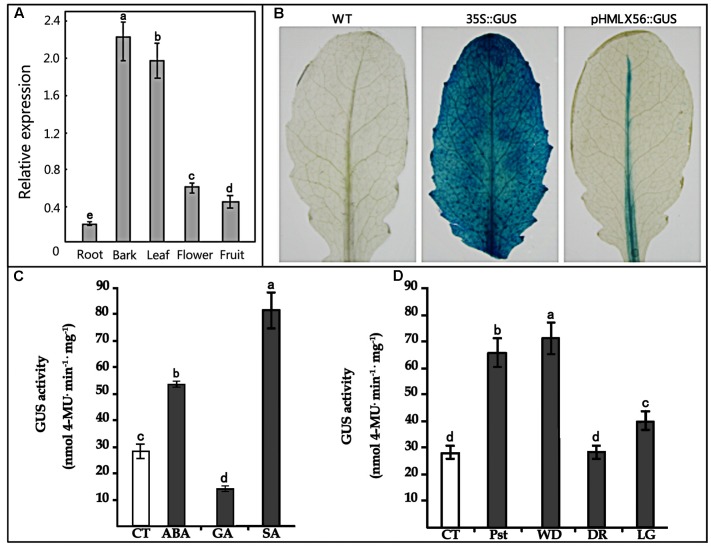
FIGURE 3.
Expression of the HMLX56 gene in different tissues. (A) mRNA expression levels of HMLX56 in mulberry tissues as analyzed by qRT-PCR. The relative gene expression was evaluated using comparative Ct method with EF1-α as the reference gene. The log2 values of the ratio of the expression of HMLX56 to EF1-α are plotted. Data are the average of three experiments for three test samples. Error bars represent SD. (B) Stable expression of pHMLX56::GUS in Arabidopsis plants. The expression vector pHMLX56::GUS was constructed by cloning the promoter of HMLX56 into the vector pBI121 to replace the cauliflower mosaic virus (CaMV) 35S promoter and drive expression of the GUS (β-glucuronidase) reporter gene. The plasmid pBI121 containing 35S::GUS was used as a positive control, and wild-type Arabidopsis plants were used as negative controls. (C,D) GUS activity driven by the pHMLX56 promoter in transgenic plants as measured by spectrophotometer. Assays were performed three times, each time with three replicates. Values are given as the mean ± SD of three experiments in each group. Different letters above the columns indicate significant differences (P < 0.05) according to Duncan’s multiple range test. WT, wild type; CT, control; Pst, Pst DC3000 infection; WD, wound treatment; DR, dark treatment; LG, light treatment.

