FIGURE 2.
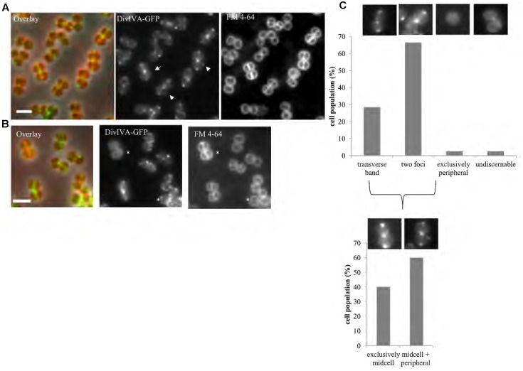
DivIVA localizes to the division site and cell periphery. Representative images of cells expressing a single copy of divIVA-gfp (A; SA247 [PdivIV A divIVA-gfp::Pspac divIVA, pGL485]). White arrows indicate cells which show both localization of DivIVA-GFP at septal membranes while also forming foci or arcs at the non-septal cell periphery. A small percentage of cells (∼2%) show only non-septal localization of DivIVA-GFP as indicated by an asterisk in (B). Live cell wide-field microscopy was performed by growing SA247 cells in BHI broth at 37°C to mid exponential phase, staining with FM 4-64 to identify septal membranes and visualized on 2% agarose pads. Scale bar represents 2 μm. (C) Histograms showing the frequency of observed DivIVA-GFP localization patterns, with example microscopic images of DivIVA-GFP localization patterns shown above the corresponding phenotypic category (n > 100 cells from 2 independent experiments). Analysis was limited to cells that showed visible septal FM 4-64 staining to exclude cells in which the septa were oriented in the x-y (lateral) plane. Analysis of cells showing septal DivIVA-GFP localization (either as a transverse band or two foci) showed that a subpopulation also displayed additional peripheral localization, as indicated in the bottom histogram and images.

