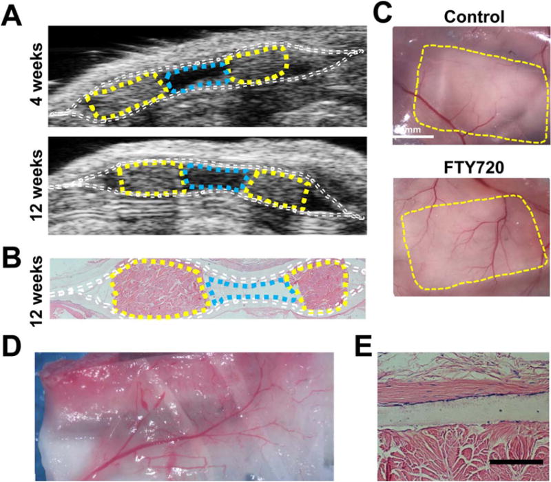
FIGURE 5.

Maintenance of implant structure imaged by ultrasound with vascularization at 12 weeks after abdominal subcutaneous implantation. (A) A subcutaneously-placed pocket monitored by ultrasound maintains a void at 4 and 12 weeks, (B) corresponding with H&E (Dotted lines: white, nanofibers; yellow, porcine decellularized dermis, cyan, void). (C) Vasculature in the intraperitoneal wall was visualized by reflection of the skin, fibers and wall upon sacrifice to compare unloaded fibers verses FTY720-loaded fibers (yellow lines: pocket edge). (D) Vascularized tissue attached to the outside of the FTY720-loaded nanofiber pocket after implantation and integration in the peritoneal cavity. (E) Histological (H&E) section of an FTY720-loaded membrane next to the structural support showing a lack of vessel penetration, scale bar 100 μm.
