Figure 6.
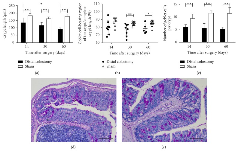
Crypt length and goblet cells. Crypt length and goblet cell numbers were determined on paraffin sections of the rectum after periodic acid-Schiff staining (PAS). (a) Crypt length was significantly decreased in the rectum of the DC group animals. (b) The length of the goblet cell-bearing region of the crypt was measured and set in ratio to the full crypt length. Thirty and sixty days postoperatively, the percentage of crypt length bearing goblet cells was decreased in the DC group. (c) Goblet cell numbers were significantly reduced in DC animals compared to the sham group. Graphs in (a)–(c) show the mean and standard error of 5 to 9 animals per group (DC 14 and 30 days, sham 14 days: n = 8; DC 60 days: n = 5; sham 30 and 60 days: n = 9); ∗p < 0.05; ∗∗p < 0.01. (d) and (e) Representative examples of sections of the rectum of DC (d) and sham (e) animals 30 days postoperatively stained with periodic acid Schiff. Crypt length (#) and goblet cells (→) can easily be identified. Scale bars represent 100 μm.

