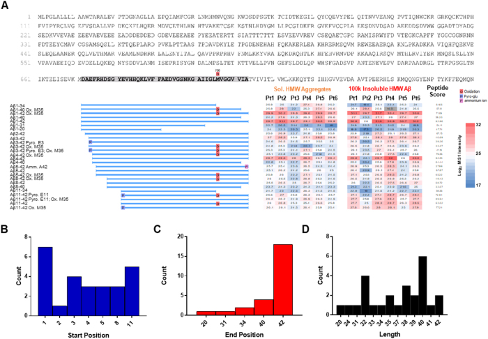
Figure 2.
Heterogeneity of Aβ proteoforms in the human Alzheimer’s disease brain. (A) Top: full protein sequence of amyloid precursor protein with Aβ1–42 sequence highlighted (grey). Left: Identified Aβ proteoform sequence names, with blue bars representing the alignment with canonical Aβ. Right: Heatmap and quantification of the log2 relative intensity of each Aβ proteoform for each of the six severe Alzheimer’s disease brain samples in the soluble aggregate and more insoluble fractions. Far right: Peptide score, indicated confidence of peptide identification38. (B) Frequency of Aβ proteoform amino acid start positions identified in CDR3 cohort; 1 = D, aspartic acid. (C) Frequency of Aβ proteoform amino acid end positions identified in CDR3 cohort; 42 = A, alanine. (D) Frequency of Aβ proteoform lengths identified in CDR3 cohort. Pt., Participant; HMW, high molecular weight; N.D., not detected; Ox., Oxidation; Pyro., Pyro-glutamate; Amm., ammonium ion.

