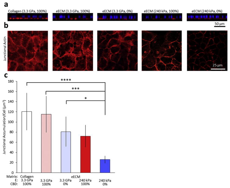
Fig. 4.
Representative immunostaining of mature Caco-2 monolayers. (a) Orthogonal sections of Caco-2 monolayers on collagen and representative eECMs, stained with phalloidin (red) and DAPI (blue), demonstrate the maintenance of proper cell polarity on all substrates. (b) Confocal micrographs of immunostained samples for junctional actin (red) and with DAPI nuclear counterstain (blue). (c) Quantification of confocal micrographs show less junctional actin accumulation on eECM with decreased CBD density and decreased stiffness. *p < 0.05, **p < 0.01. (For interpretation of the references to colour in this figure legend, the reader is referred to the web version of this article.)

