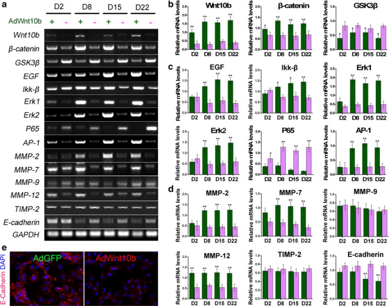
Fig. 5.

Wnt10b activated gene expressions of Wnt, EGF, and MAPK pathways. a–d RT-PCR and bar charts revealed significant increase in Wnt10b, β-catenin, EGF, Ikk-β, Erk1, Erk2, AP-1, MMP-2, 7, 12 and dramatic decrease in Gsk3β, p65, and E-cadherin at different time points in AdWnt10b-treated groups when compared to those of the AdGFP-treated groups. e Immunostaining showed E-cadherin expression was decreased in AdWnt10b-treated cells compared with the AdGFP-treated group. n > 5, *p < 0.05, **p < 0.01
