Figure 1.
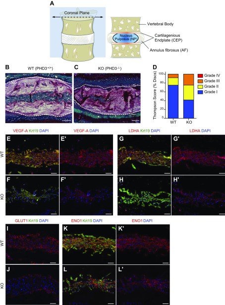
PHD3−/− mice show increased incidence of disc degeneration and decreased expression of select HIF-1 target genes in NP tissue. A) Schematic drawing of spinal motion segment and coronal cross section showing vertebral bodies and the intervertebral disc with its central NP, circumferential AF, and superior and inferior cartilaginous endplates (CEP). B, C) Representative Safranin-O/Fast Green/hematoxylin–stained sections of 12.5-mo-old WT (B) and PHD3−/− (C) mice. The PHD3−/− mouse showed a decreased number of NP cells and some changes in cell morphology. D) PHD3−/− mice showed increased incidence of discs with a higher grade of degeneration, as assessed by the Thompson grading scale. Four discs/animal were scored from 3 pairs of littermate animals. E–L') Representative immunofluorescence images from 12.5-mo-old WT (PHD3+/+) and knockout (PHD3−/−) mice showing NP tissue areas with a comparable number of cells. There was decreased staining of HIF-1 targets VEGF-A (E–F'), LDHA (G–H'), and GLUT1 (I, J) in knockout mice compared to WT mice. In contrast, another HIF-1 target, ENO1, shows comparable expression in PHD3+/+ (K, K') and PHD3−/− (L, L') mice. Krt19 was used as a marker to label the NP tissue compartment with VEGF-A, LDHA, and ENO1 (independent channel not shown). Scale bars, 100 μm (B, C) and 50 μm (E–L'). Images are representative from 3 independent littermate groups.

