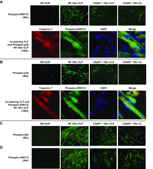
Figure 2.
Phospho-MAPKs and Akt before (Ctrl, control) and 16 h after CLP, in frozen LV sections of WT mouse hearts or C5aR-KO mouse hearts. A) Phospho-ERK-1/2 before (Ctrl) or 16 h after CLP. Red: TnT; green: phospho-ERK-1/2; blue: DAPI-stained nuclei; yellow-green: merged image of TnT and ERK-1/2 labeling in CMs. A) Top: phospo-ERK-1/2 revealed green staining of CMs 16 h after CLP. The staining was markedly reduced in CLP CMs from C5aR-KO mice. B) Phospho-p38 images were very similar to those for phospho-ERK-1/2. C, D) Phospho-Akt (C) and phospho-JNK-1/2 (D) 16 h after CLP in WT CMs and CMs with and without C5aR1 and -R2. There were marked reductions in phospho-Akt and phospho-JNK-1/2 in frozen LV sections from C5aR-KO mice (n ≥ 3 for each group of frozen sections).

