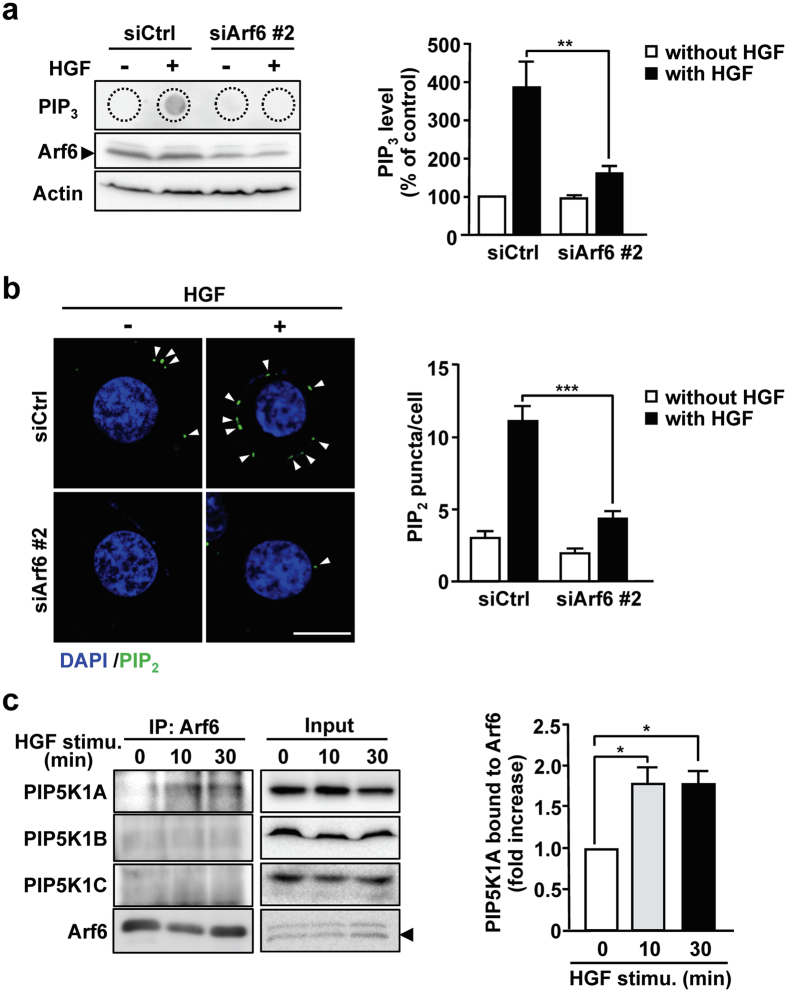
Figure 2.
Arf6 promotes PIP2 and PIP3 production by activating PIP5K1A upon HGF stimulation of HepG2 cells. (a) HepG2 cells transfected with siRNAs for control and Arf6 were stimulated with HGF as in Fig. 1a. After lipids were extracted from the cells, PIP3 was detected by dot blotting with anti-PIP3 antibody (left panels). Arf6 and actin in cell lysates were also detected by Western blotting. PIP3 levels were quantified (right panel). (b) HepG2 cells transfected with siRNAs for control and Arf6 were stimulated with HGF as in Fig. 1a, and immunostained for PIP2 (green) and stained for nuclei with DAPI (blue) (left panels). The number of PIP2 puncta shown by arrow heads in the images was counted and normalized by cell number (right panel). Scale bar, 10 μm. (c) HepG2 cells were stimulated with 10 ng/mL of HGF for the indicated time. Endogenous Arf6 was immunoprecipitated, and co-precipitated PIP5K1 isoforms were detected by Western blotting with antibodies specific to each PIP5K1 isoform (left panels), and co-precipitated PIP5K1A was quantified (right panel). All quantitative results represent means ± SEM from at least three independent experiments. Statistical analyses: two-way ANOVA with post hoc Bonferroni’s test (a and b) and one-way ANOVA with post hoc Tukey’s test (c). *p < 0.05; **p < 0.01; ***p < 0.001. Blots shown in (a and c) are cropped images. The full-length blots are presented in Supplementary Figure.

