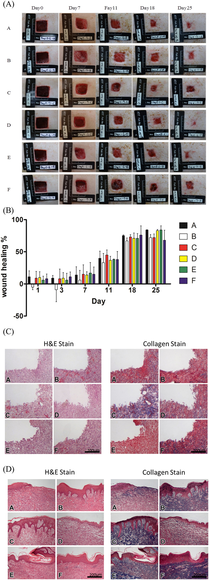
Figure 6.

Diabetes mellitus was induced in Landrace pigs, and 2 × 2-cm wounds were created to observe wound healing. (A) The wound area size of the original type, original type mixed with 5% collagen, original type mixed with 350-ppm silver nanowire, original type mixed with 175-ppm silver nanowire, original type mixed with 5% collagen, 5% chitosan, and 350-ppm silver nanowire, and original type mixed with 5% collagen, 5% chitosan, and 175-ppm silver nanowire at day 0, day 7, day 11, day 18 and day 25. (B) The wound healing area quantitative analysis. (C) A 10th-day tissue section stained with H&E stain and Masson’s trichrome stain. The scale bar is 200 micrometer (D) A 21st-day tissue section stained with H&E stain and Masson’s trichrome stain. The scale bar is 200 micrometer.
