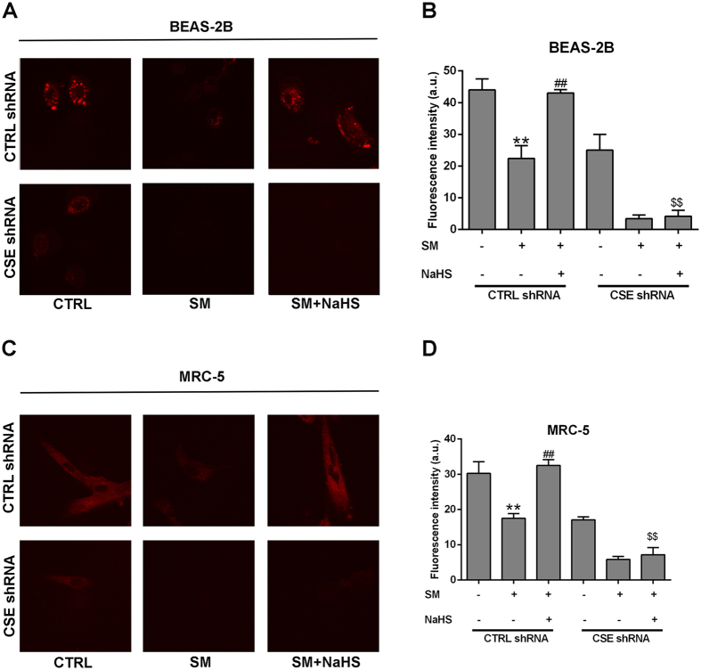
Figure 7.
Confocal fluorescence imaging of endogenous H2S in living BEAS-2B and MRC-5 cells using NIR-HS. Untreated or SM-treated BEAS-2B and MRC-5 cells were further cultured for 24 h. BEAS-2B Cells were incubated with NIR-HS (5 μM) for 10 min (A). MRC-5 Cells were incubated with NIR-HS (5 μM) for 10 min (C). The average fluorescence intensity of cells in above images (G). Data are presented as the mean ± SEM (n = 5). **p < 0.01 vs A column; ##p < 0.01 vs B column; $$p < 0.05 vs C column.

