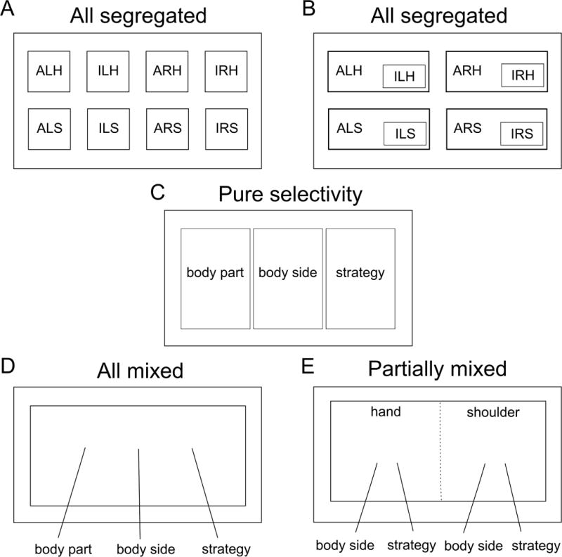
Figure 3. Possible organizational models of neural representations.

(A) The neurons coding for each condition are anatomically segregated, i.e. distinct, non-overlapping networks. (ALH = Attempt Left Hand, ILH = Imagine Left Hand, ARH = Attempt Right Hand, IRH = Imagine Right Hand, ALS = Attempt Left Shoulder, ILS = Imagine Left Shoulder, ARS = Attempt Right Shoulder, IRS = Imagine Right Shoulder). (B) Conditions can be overlapping such that the responses to some conditions are subsets or weak versions of others, e.g. imagined movements being subsets of attempted movements. (C) Neurons coding each of the motor variables (body part, body side, and strategy) are anatomically segregated. (D) The neural population exhibits mixed selectivity, with individual neurons showing tuning to various conjunctions of variables. (E) The neural population exhibits partially mixed selectivity, with the mixing of representations being dependent on the variables under investigation. Here, hand and shoulder are mixed leading to orthogonal coding of effectors (functional segregation), however, the other variables (body side and strategy) are mixed only within, but not between, effectors. This model is consistent with the results observed in this study. Note that solid lines in this diagram indicate anatomical boundaries of neural populations while the dotted line indicates functional boundaries/segregation.
