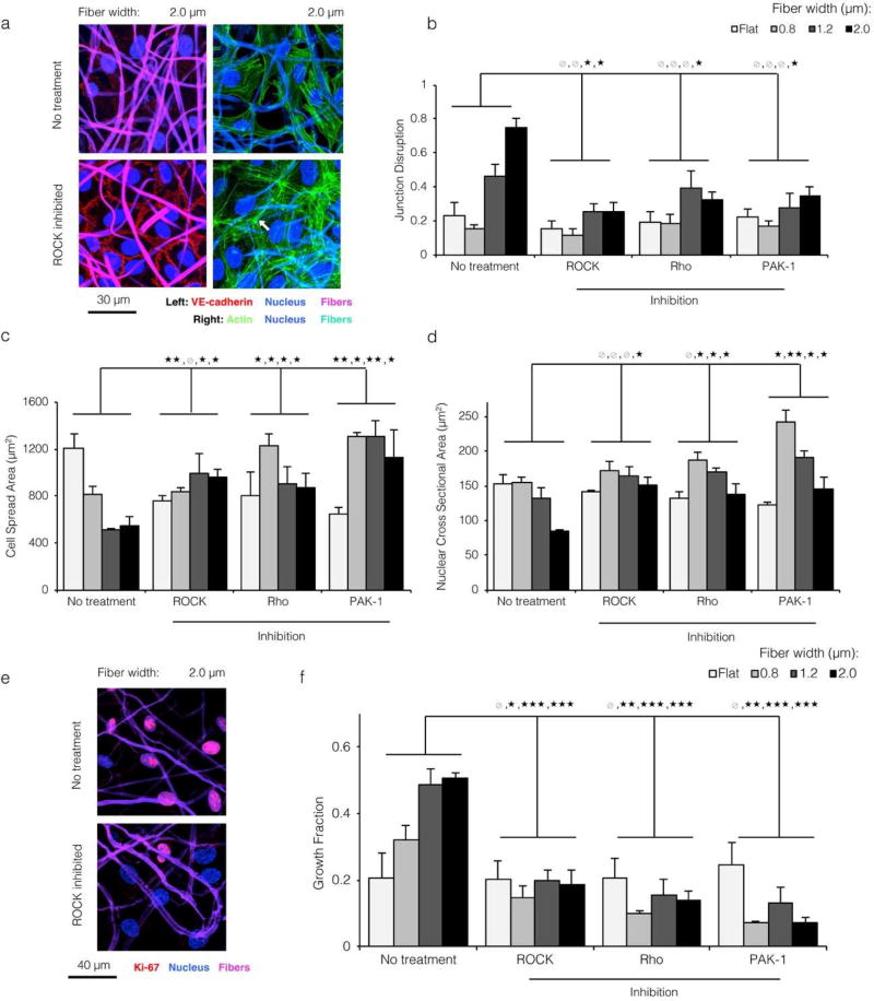
Figure 4. Topographical cues modulating EC morphology and proliferation are dependent on Rho, ROCK, and Rac signaling.
a) ECs cultured on the most topographically varied substrate (2.0 µ wide fibers), treated with ROCK inhibitor Y-27632, and stained for the nucleus (blue) and VE-cadherin (red) or actin (green). Fibers are shown in violet (left panels) or cyan (right panels). Chemical inhibition of cytoskeletal signaling prevented both the loss of VE-cadherin junctions and the reorganization of actin away from junctions (white arrow), despite the underlying substrate topography. b,c,d) For all topographical conditions, chemical inhibition of Rho, ROCK, and Rac signaling led to EC morphology reminiscent of that on flat and small width fibrous substrates: low junction disruption (b), large planar spreading area (c), and large nuclear area (d). This suggested that the morphological shifts associated with more varied substrate topography were dependent on cytoskeletal signaling. e,f) ECs cultured on large width ELP fibers and treated with cytoskeletal inhibitors proliferated at a low level, consistent with a regain of contact inhibition by VE-cadherin. (* p < 0.05, ** p < 0.01, *** p < 0.001)

