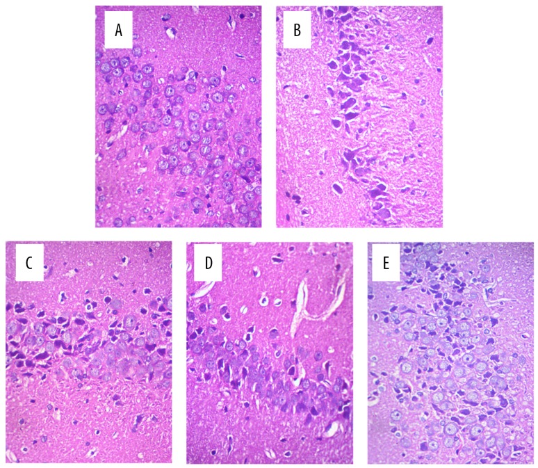
Figure 2.
Microscopic observation of nerve cells in the hippocampus of rats after MCAO (HE staining, 400×). (A) Hippocampal neurons in the sham group. The neurons in the hippocampus were in neat rows, close together, and of normal cell morphology and size. (B) Hippocampal neurons in the model group. The neurons in the hippocampus were arranged loosely, with cell body shrinkage, partial nuclear pyknosis, nuclear fragmentation, and nucleolar blurring and even disappearance. (C) Hippocampal neurons in the NaB group. (D) Hippocampal neurons in the 20 mg/kg apigenin group. (E) Hippocampal neurons in the 40 mg/kg apigenin group. As shown in panels (C–E), the hippocampal neuron damage was alleviated after ischemia and reperfusion injury in the three intervention groups.

