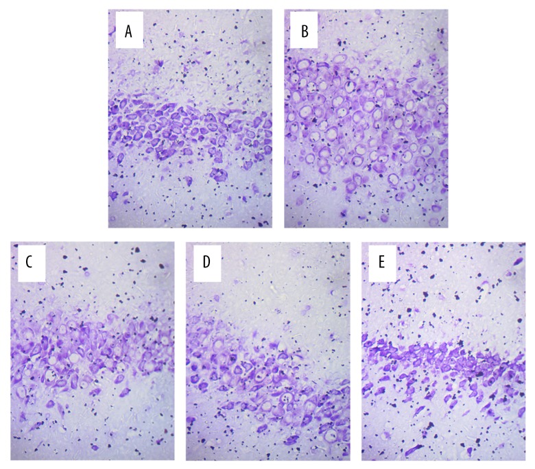
Figure 3.
Microscopic observation of nerve cells in the hippocampus of rats after MCAO (Nissl staining, 400×). (A) Hippocampal neurons in the sham group. The Nissl staining showed that the Nissl bodies of nerve cells were blue, and that they were distributed around the nucleus in an orderly manner, with complete and normal cellular structure and clear nuclei. (B) Hippocampal neurons in the model group. The Nissl bodies in the model group were released, the cyton of the neurons was shrunk, there was neuronal degeneration, and the neurons were decreased. (C) Hippocampal neurons in the NaB group. (D) Hippocampal neurons in the 20 mg/kg apigenin group. (E) Hippocampal neurons in the 40 mg/kg apigenin group. As shown in panels (C–E), the hippocampal neuron damage was alleviated after ischemia and reperfusion injury in the three intervention groups.

