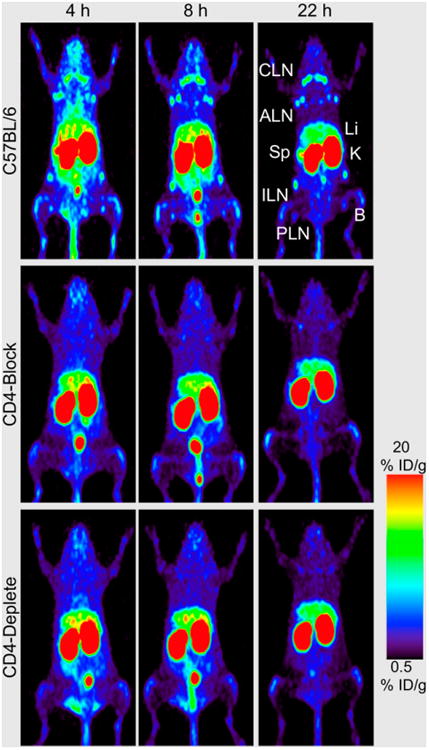
Figure 1.

Anti-CD4 small-animal PET imaging (using 89Zr-malDFO-GK1.5 cys-diabodies, where malDFO is N-(3,11,14,22,25,33-hexaoxo-4,10,15,21,26, 32-hexaaaza-10,21,32-trihydroxytetratriacontane) maleimide) of T-lymphocytes in C57BL/6 wild-type, CD4-blocked, and CD4-depleted mice at 4, 8, and 22 h after injection. Images are 25-mm maximum-intensity projections. Compared with wild-type mice, both CD4-blocked and CD4-depleted animals lack uptake in axillary lymph nodes (ALN), cervical lymph nodes (CLN), inguinal lymph nodes (ILN), popliteal lymph nodes (PLN), and spleen (Sp). B 5 bone; ID 5 injected dose; K 5 kidney; Li 5 liver. (Adapted from (11).)
