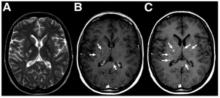
Figure 2.

Axial MR images of inflammation in brain of young woman with multiple sclerosis. (A) Unenhanced T2-weighted image shows multiple hyperintense lesions. (B) Gadolinium-enhanced T1-weighted image before injection of USPIO shows 3 enhancing lesions (arrows). (C) At 24–48 h after injection, the original 3 lesions are seen along with 3 additional lesions (arrows). (Adapted with permission of (20).)
