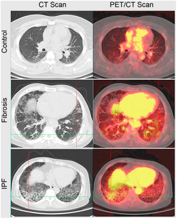
Figure 4.

PET/CT imaging of lung inflammation. 68Ga-BMV101 PET/CT probe (targeting cysteine cathepsins, which are highly expressed in activated macrophages) was injected intravenously, and images were collected 2.5 h later. Patient with idiopathic pulmonary fibrosis (IPF) shows increased accumulation in fibrotic regions, but patient with unclassified fibrosis shows no significant increase compared with normal-lung control. (Adapted with permission of (35).)
