Figure 3.

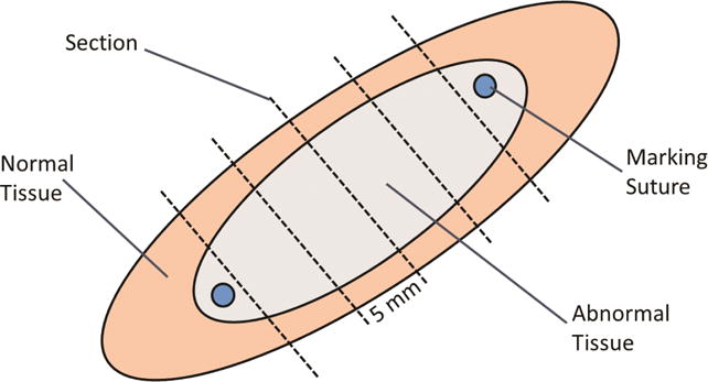
Schematic diagram of section preparation. Each lesion was excised with a rim of normal tissue surrounding it. Sections were taken transversely every 5 mm along the lesion.

Schematic diagram of section preparation. Each lesion was excised with a rim of normal tissue surrounding it. Sections were taken transversely every 5 mm along the lesion.