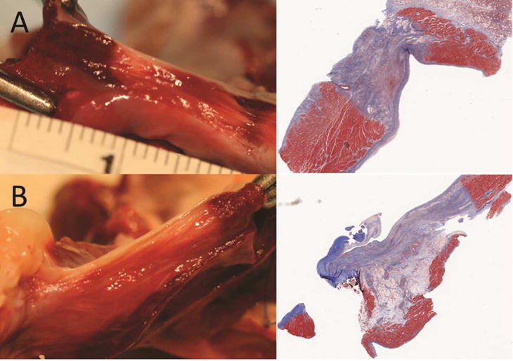
Figure 6.

Representative transmural (A) and non-transmural (B) sections by TTC (left) and Masson’s trichrome (right). Viable tissue stains dark red with TTC; nonviable tissue appears yellow to white. Collagen in scar tissue stains blue with Masson’s trichrome; muscle tissue stains red.
