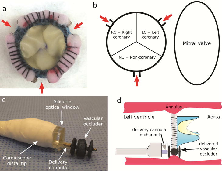
Fig. 2.

Paravalvular leak model and device deployment. (a) Bioprosthetic valve incorporating 3 PVLs. (b) Schematic showing PVL locations as viewed from ventricle. (c) Three-lobe occluder (Amplatzer AVP II) shown extended from delivery cannula as deployed from cardioscope. (d) Delivery process.
