Fig. 5.

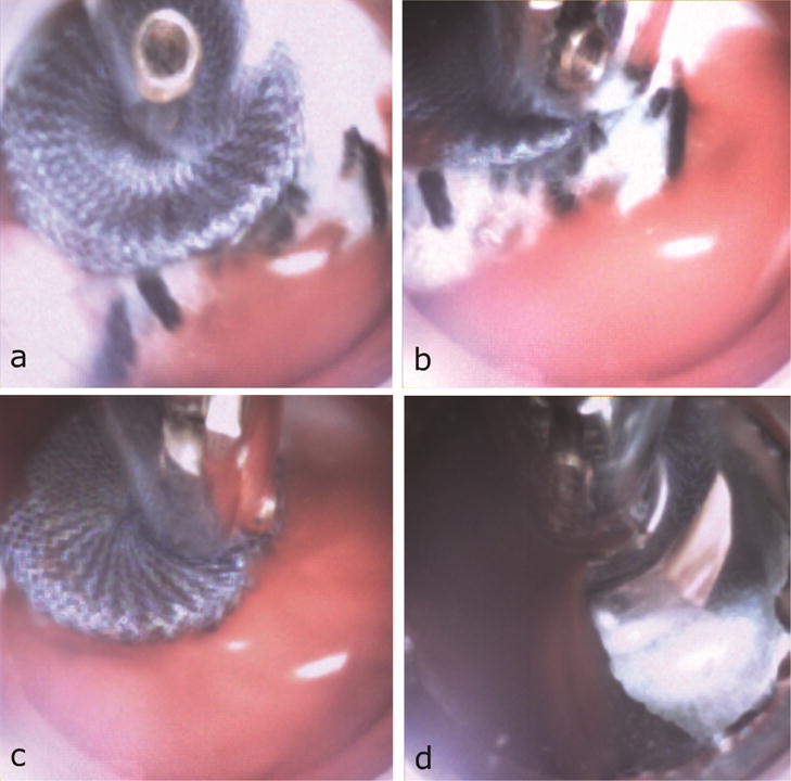
Cardioscopic images during device recovery. (a) Positioning device screw connector inside working channel. (b) Forceps grasping screw connector. (c) Device pulled from PVL. (d) Device removed through apex.

Cardioscopic images during device recovery. (a) Positioning device screw connector inside working channel. (b) Forceps grasping screw connector. (c) Device pulled from PVL. (d) Device removed through apex.