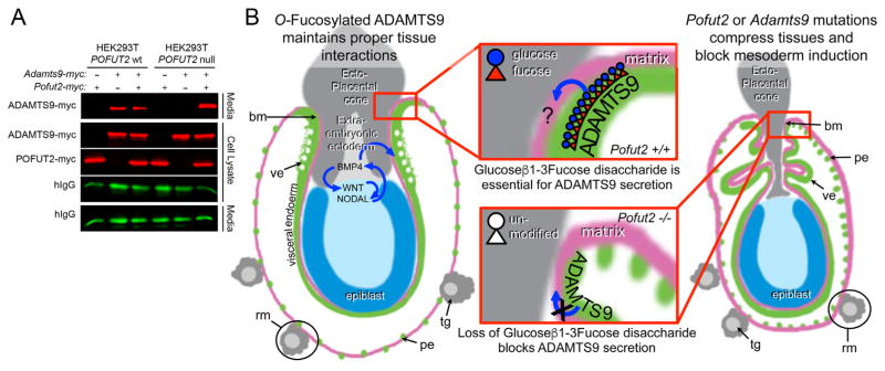
Fig. 6.
Loss of POFUT2 blocks secretion of ADAMTS9 in HEK293T cells.(A) Western blot analysis of myc-tagged ADAMTS9-N-L2 (ADAMTS9-myc) secretion from wild-type HEK293T cells or Pofut2 CRISPR/Cas9-targeted HEK293T cells (HEK293T Pofut2 null). Cells were co-transfected with expression plasmids encoding Adamts9-myc and hIgG with or without additional Pofut2-myc or empty vector. In HEK293T cells, ADAMTS9 is secreted into the media with or without transfected Pofut2-myc. In contrast, in CRISPR/Cas9-mutated HEK293T cells ADAMTS9, although observed in the cell lysate, fails to be secreted into the medium. Co-transfection with Pofut2-myc rescues secretion defects in HEK293T Pofut2 null cells. (B) Model for action of POFUT2 and ADAMTS9 in the early gastrula. We propose that O-fucosylation of ADAMTS9 promotes the proper arrangement of extraembryonic tissues (grey and green) critical for specification of visceral endoderm characteristics (green) and maintaining signals essential for mesoderm induction in the epiblast (blue). We hypothesize that one possible function of ADAMTS9 is to act locally (red square) to anchor proximal visceral endoderm cells to the ECM or ectoplacental cone cells. We predict that loss of Pofut2 (by blocking secretion of ADAMTS9) or loss of Adamts9 disrupts this anchor point causing the visceral/parietal endoderm layers to slip. This disturbance alters the properties of the extraembryonic ectoderm or displaces the tissue. As a consequence, the visceral endoderm does not receive BMP signals essential for maintaining characteristics such as apical vacuoles (white spots) and the epiblast is unable to maintain signals needed for mesoderm induction (WNT and NODAL). The increased laminin staining in Reichert’s membrane (pink) in Pofut2 mutants (Du et al., 2010) could result from loss of this anchor point (retraction of the membrane), or alternatively from loss of ADAMTS9 function in the parietal endoderm cells or trophoblast giant cells. Wild-type and mutant embryos are oriented proximal up and distal down. Abbreviations: ectoplacental cone (ec), epiblast (ep), parietal endoderm (pe), Reichert’s membrane (rm), trophoblast giant cells (tg), visceral endoderm (ve).

