
Fig. 5.
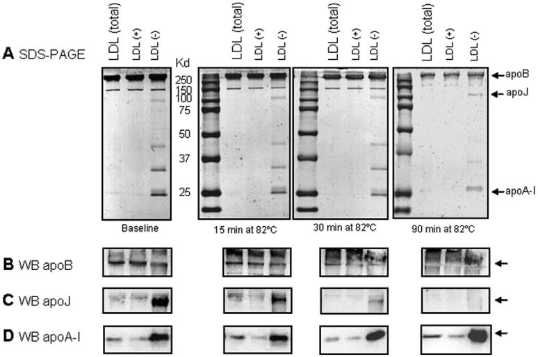
Protein degradation upon thermal denaturation of LDL. (A) SDS-PAGE (10%, Coomassie blue staining) was used to assess changes to apoB after 15, 30 and 90 min of incubation at 82 °C. Gels were run at 100 V for 1.5 h. (B, C) Following SDS-PAGE, Western blot analysis was used to detect apoB (B), apoJ (C) and apoA-I (D).