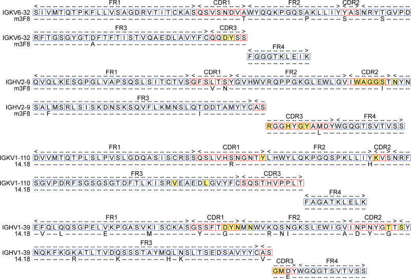
Figure 2.
Alignment of anti-GD2 antibodies with their putative germline sequences. Top rows representative of germline amino acid structure. Bottom rows represent mutations from the germline antibody (dashed = no mutation). Amino acids relevant in antigen binding, as proposed by Ahmed et al. (2013), are highlighted in yellow. Complementary determining regions (CDRs) are labeled as designated by IMGT analyses.

