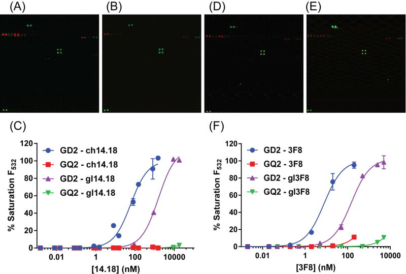
Figure 3.
High specificity of anti-GD2 antibodies for antigen GD2. (A) Affinity mature 14.18 and (B) germline 14.18 antibody react with high specificity towards antigen GD2. (C) The non-target antigen with the highest observable binding was GQ2, at greater than 500-fold reduced levels. (D) Affinity mature 3F8 and (E) germline 3F8 were also extremely specific for GD2. (F) GQ2 binding was observed in 3F8 at levels greater than 250-fold reduced versus that of GD2. Linear formats of these same data are demonstrated in Figure S3 to fully illustrate antibody dose saturation. Data are represented as mean ± standard deviation (minimum of 2 independent array experiments, duplicate spots per array). See also Figures S1–S5 and Table S2.

