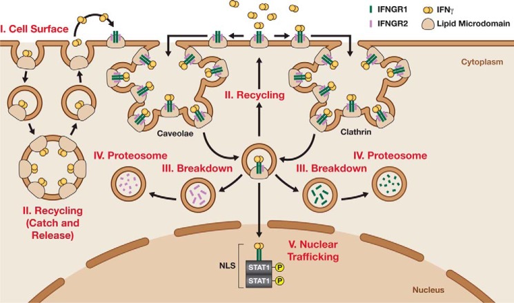
Figure 2.
Endocytic mechanisms regulating IFNγ signaling via differential expression of the IFNGR. IFNGR1 (green) and IFNGR2 (purple) are associated in lipid micro-domains on the cell surface. In the presence of IFNγ dimers (orange), the cytokine receptor complex is redirected to clathrin or caveolae sites at the cell surface. Once there, IFNγ either binds the receptor or is recruited to PS-rich domains. Then, the IFNG complexes are endocytosed either through clathrin-dependent events or clathrin-independent events that involve caveolae-dependent mechanisms. IFNγ–PS complexes are slowly recycled back to the cell surface, and IFNγ is released in an autocrine-like manner (left). In contrast, IFNγ–IFNGR complexes are endocytosed and broke down before being recycled or degraded. Noticeably, IFNGR2 is enzymatically cleaved and separated for degradation via proteasomes. Meanwhile, IFNGR1 can be recycled to the cell surface or targeted for degradation. Alternatively, the IFNG–IFNGR–p-STAT1 complex could remain stable and continue translocating to the nucleus using NLS.

