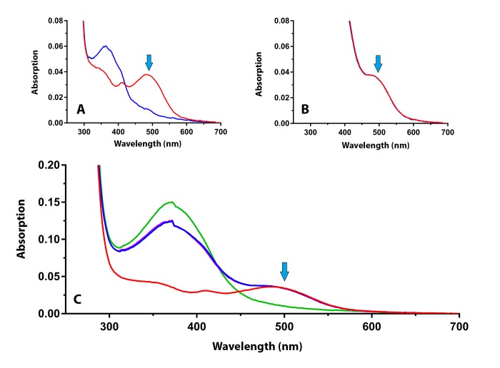
FIGURE 7.
Spectral scans of the retinal extracts from the donor eye with advanced macular degeneration shown in Figure 6. (A) The rhodopsin spectrum before (red line) and after (blue line) bleaching with bright light. (B) The dark-adapted spectrum after the addition of exogenous 11-cis retinal (2 min red line; 20 min blue line) to test for rhodopsin regeneration. (C) The full spectral curves before and after the addition of 11-cis retinal, and after bleaching in bright light. No increase in rhodopsin absorption is detected at 498 nm (blue arrows).

