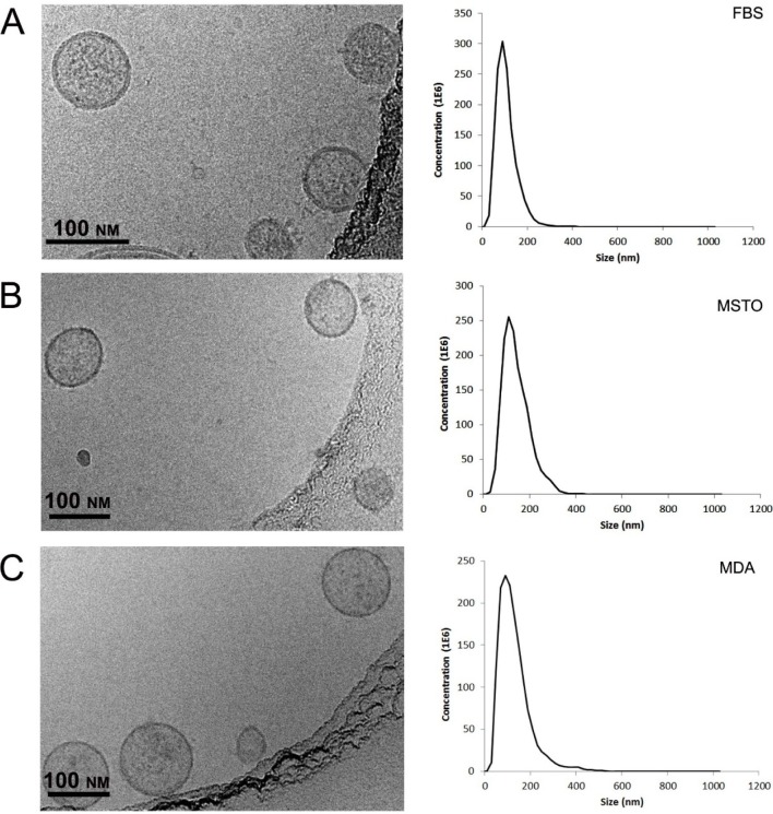
Figure 2.
Characterization of purified extracellular vesicles isolated from FBS (A), MSTO cells (B), and MDA cells (C). Representative Cryogenic Transmission Electron images of vesicles show round-shape membrane-bound particles from all three sources (FBS, MSTO and MDA cells; left column) with occasional irregularly distributed electron-dense content, both inside and on the surface of the membrane. The NanoSight plots depict typical size distributions of extracellular vesicles from the above-described sources (right column). Bars correspond to 100 nm.

