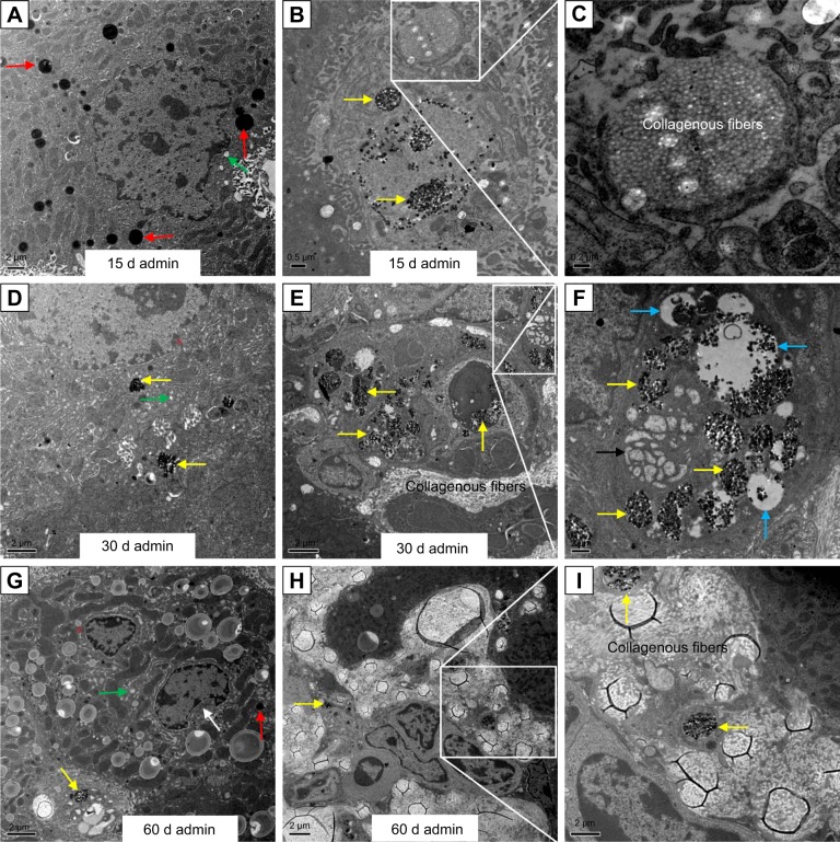
Figure 6.
Ultrastructural changes in the liver of the SiO2 NP treated mice at days 15, 30, and 60.
Notes: (A) Nuclear membrane distorted (white star), vacuolization (green arrows), and lipid droplet formation (red arrows) in the hepatocytes induced by the SiO2 NPs in the liver at day 15 (5 K). (B) SiO2 NPs (yellow arrows) in the hepatic sinusoid with proliferation of the collagen fibers starting at day 15 (8 K). (C) Magnification of the collagen fibers (25 K). (D) Nuclear membrane indentation with chromatin margination (red stars) and vacuolization (green arrows) in the hepatocytes in the liver at day 30 (8 K). (E) Hepatic granuloma mainly consisted of different kinds of cells at day 30 (4 K), (F) lamellar body-like structure formation (black arrow) and vacuolization (blue arrows) in the granuloma at day 30 (10 K). (G) Chromatin margination (red star), crescent-shaped nucleus (white arrow), and lipid droplet formation (red arrow) in the cytoplasm as well as vacuolization (green arrow) in the hepatocytes in the liver at day 60 (2.5 K). (H) Fibrotic granuloma induced by SiO2 NPs at day 60 (3 K). (I) Magnification of the SiO2 NPs surrounded by the white collagen fibers (8 K).
Abbreviations: admin, administration; SiO2 NPs, silica nanoparticles.

