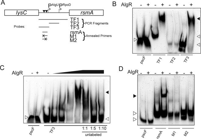
FIG 4.
AlgR directly binds the AlgU-dependent rsmA promoter and requires both AlgR-binding sites. Purified AlgR was incubated with either biotinylated PCR products or annealed primers. (A). Schematic of the rsmA genomic region. Below are fragments or annealed primers and the approximate location upstream of rsmA. Promoters are indicated above as bent arrows and are denoted by PAlgU or PRpoD. The arrowheads indicate the approximate location of the putative AlgR-binding sites. (B) Analysis of biotinylated PCR fragments in gel shift studies. The PCR fragment TF1 represents the entire rsmA upstream region (see panel A). Fragments TF2 and TF3 correspond to the separated rsmA promoters (see Fig. 1). TF2 corresponds to the proximal rsmA promoter, and TF3 corresponds to the distal AlgU-dependent promoter. A minus sign indicates probe alone. A plus sign indicates AlgR concentration of 2.5 μM. pscF was used as a negative control. Shaded arrowheads indicate shifted fragments. White arrowheads indicate unbound probe. (C) Titration and competition assay using purified AlgR and the TF3 fragment corresponding to the distal rsmA promoter. The pscF fragment was used as a negative control. TF3 was incubated with different concentrations of AlgR (indicated by the graded triangle; 0.05, 0.25, 1.25, and 2.5 μM). TF3 was also used in a competition assay using unlabeled TF3 as a competitive inhibitor. Ratios below indicate the ratio of labeled to unlabeled probe. Shaded arrowheads indicate shifted fragments. White arrowheads indicate unbound probe. A minus sign indicates probe alone. (D) Two AlgR-binding sites are upstream of the distal rsmA promoter. Annealed primers containing either wild-type or two different mutated AlgR-binding sites were tested in gel shift experiments. The pscF fragment was used as a negative control. The rsmA lanes indicate the probe using annealed primers with both AlgR-binding sites intact. M1 indicates the same as rsmA, except that the furthest upstream AlgR-binding site was mutated. M2 indicates the same as rsmA, except that the further downstream AlgR-binding site is mutated. Shaded arrowheads indicate shifted fragments. White arrowheads indicate unbound probe. Minus signs indicate probe alone. Plus signs indicate an AlgR concentration of 2.5 μM.

