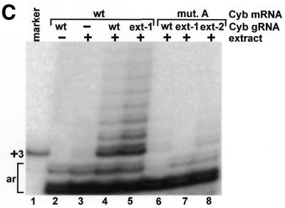
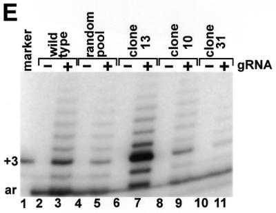
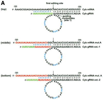
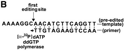
Figure 1.


gRNA-binding is not sufficient for editing. (A) Base-pairing of a cytochrome b (Cyb) gRNA and mRNA transcript. A poly(U) tail that is added to the 3′ end of the gRNA by a TUTase activity within the editing extract (green) has the potential to interact (blue) with the purine-rich pre-edited sequence. Mutations (mut.) to the mRNA that would disrupt this interaction and two compensatory extensions (ext.) to the 3′ end of the gRNA are shown in red. The A→C substitutions within the gRNA that stimulate the in vitro reaction are highlighted in blue and the guiding nucleotides are in lower case. The first editing site and the corresponding guiding nucleotides are indicated. (B) The primer extension reaction used to detect in vitro U-insertions within the first editing site. A primer annealed immediately 3′ of the first cytochrome b editing site is extended by a number of radiolabeled dA nucleotides corresponding to the number of U nucleotides inserted during editing. Addition of ddG terminates the reaction. (C) Cytochrome b mRNA transcripts containing the wild-type and mutated pre-edited sequence were assayed for gRNA-directed U-insertions using no added gRNA (–), and gRNAs with and without (wt) a compensatory 3′ extension. The marker for correct editing results from the extension of a template containing three insertions at the first editing site (+3). Additional bands are artifacts of the assay (ar) as indicated by their presence in the non-extract treated lanes. (D) The wild-type cytochrome b editing domain and the sequence of three RNAs cloned from the random RNA pool. The sequence that was made random is shown in square brackets. (E) Assay of the above RNAs for gRNA-directed U-insertions within the first editing site.


