Figure 5.
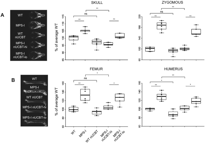
Neonatal UCBT prevents bone thickening in MPS-I mice. (A) On the left, representative radiographs of the skull of 20-weeks-old WT, MPS-I, WT nUCBT, MPS-I nUCBT-hi, and MPS-I nUCBT-lo mice. On the right, measurements of the skull width and zygomous width, performed on radiographs of WT (n = 6, 3 males and 3 females), MPS-I (n = 6, 3 males and 3 females), WT nUCBT (n = 7, 3 males and 4 females), MPS-I nUCBT-hi (n = 5, 3 males and 2 females), and MPS-I nUCBT-lo mice (n = 5, 3 males and 2 females). (B) On the left, representative radiographs of the femur of 20-weeks-old WT, MPS-I, WT nUCBT, MPS-I nUCBT-hi, and MPS-I nUCBT-lo mice. The increase in meta-diaphyseal bone density observed in MPS-I (asterisks) is significantly prevented in MPS-I nUCBT-hi mice. On the right, measurements of the femur and humerus widths, performed on the radiographs of the same animals as in panel A. *p ≤ 0.05, **p ≤ 0.01, by Wilcoxon test.

