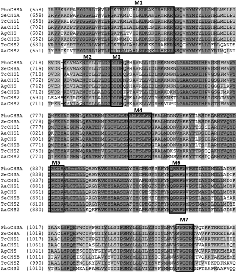
Figure 3.
Partial amino acid alignment of PhoCHSA with selected CHS enzymes from other insects showing the conserved motifs found in all known insect CHS proteins. Identical residues are shown in dark grey background while conserved residues are shown in light grey background. M1 and M2: nucleotide binding sites similar to Walker A and Walker B motifs, M3 and M4: donor saccharide binding sites. M5: Acceptor binding site, M6: product binding site, M7: involved in chitin extrusion along with the 5TMS region (not shown). Aa: Aedes aegypti, Ag: Aphis glycines, Se: Spodoptera exigua, Tc: Tribolium castaneum, AaCHS1: AAEL002718-PA, AaCHS2: AAEL005618-PA, AgCHS: AFJ00066, SeCHSA: AAZ03545, SeCHSB: ABI96087, TcCHS1: NP_001034491, TcCHS2: NP_001034492.

