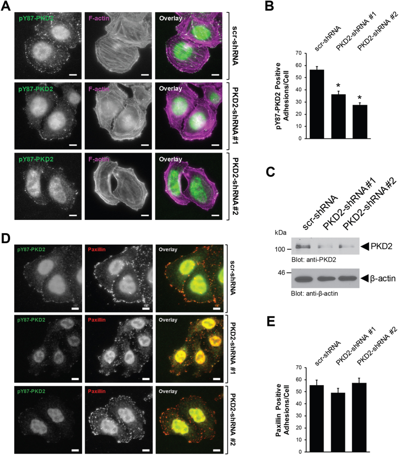
Figure 2.
The anti-pY87 signal at the focal adhesions corresponds to PKD2. (A) HeLa cells were infected with lentivirus expressing non-target shRNA (scr-shRNA), or two independent shRNA sequences targeting PKD2 (PKD2-shRNA#1, PKD2-shRNA#2). After 48 hours, cells (6000/channel) were reseeded in ibidi channel μ-slides. Cells were subjected to immunofluorescence analysis to determine the localization of endogenous pY87-PKD2 (secondary antibody used: Alexa Fluor 488) and F-actin (phalloidin). Scale bar indicates 10 μm. (B) Shows quantitation for the number of pY87-PKD2-positive adhesions per cell with scr-shRNA (n = 73 cells), PKD2-shRNA#1 (n = 63 cells) and PKD2-shRNA#2 (n = 64 cells). Error bars represent standard error of the mean (SEM). *Indicates statistical significance (p < 0.05) as compared to cells expressing scr-shRNA. (C) Cell lysates from A were evaluated by Western blotting for the expression of PKD2 and β-actin. All experiments have been performed at least three times with similar results. Uncropped blots are shown in Supplemental Figure S7. (D) HeLa cells were infected and reseeded in ibidi channel μ-slides, as described in (A) Cells were subjected to immunofluorescence analysis to determine the localization of endogenous pY87-PKD2 and paxillin. Scale bar indicates 10 μm. (E) Shows quantitation for the number of paxillin-positive adhesions per cell with scr-shRNA (n = 40 cells), PKD2-shRNA#1 (n = 40 cells) and PKD2-shRNA#2 (n = 40 cells). Error bars represent standard error of the mean (SEM). *Indicates statistical significance (p < 0.05) as compared to cells expressing scr-shRNA.

